19415 Deerfield Avenue • Suite 112 • Lansdowne, VA 20176 • 703-724-1195
1860 Town Center Drive • Suites 150 & 160 • Reston, VA 20190 • 703-480-0220
224-D Cornwall Street, N.W. • Suite 400 • Leesburg, VA 20176 • 703-443-6733
24440 Stone Springs Blvd • Suite 545 • Dulles, VA 20166 • 703-957-1022
1801 Robert Fulton Drive • Suite 510 • Reston, VA 20191 • 703-783-5355
Hematuria (Blood in the Urine)
Hematuria is a word used to refer to blood in the urine. “Gross” hematuria refers to blood that can actually be seen in the urine. The urine may appear red in color or have a “rusty” or “tea color” to it. Microscopic hematuria refers to blood which cannot be seen but which is detected on urinalysis either by a chemical dipstick test or by examination of the urine under a microscope. When hematuria is present, the urinary tract requires evaluation.
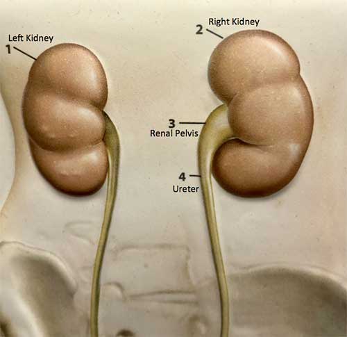
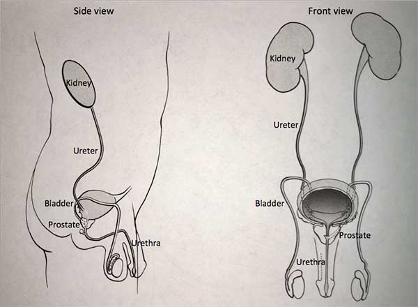
The urinary tract begins with the kidneys. The kidneys, one on each side, sit high in the upper abdomen partially underneath the rib cage.
They filter the blood to extract excess waste products and fluid to form the urine. Urine, once formed in the kidneys, travels through a tube on each side, the ureter, down to the bladder. Urine is constantly being made by the kidneys and transported through the ureters into the bladder. The bladder stores urine until full and then empties to the outside through the urethra. The urinary system is the same in both men and women from the kidneys to the bladder. In men, the urethra is longer and encircled by the prostate which is a gland that is part of the reproductive system.
When blood is present in the urine, it is often not a sign of anything which poses a significant threat to the health. As many as 10% of people may have blood in the urine. One of the most common causes for blood in the urine occurs when the kidneys first filter the blood to make the urine. There may be some blood cells which slip through the filter and end up in the urine. Another common cause of blood in the urine in both men and women is mild inflammation which may be present in the lower portion of the bladder. In men, there may be blood in the urine due to benign prostatic hyperplasia, which is the normal enlargement of the prostate that occurs in all men beginning at the age of 40.
Evaluation for Hematuria
The primary goal of evaluation is to determine if there is underlying abnormality which would put the patient’s health at risk. The urinary system is investigated to determine if there may be infection, obstruction, scarring, stone or tumor.
Evaluation for hematuria usually begins with a study of the upper part of the urinary system. Some type of x-ray, known as an imaging study, is carried out to gather information about the kidneys, the ureters, and the bladder. Imaging studies usually include either a sonogram or a CT urogram. A sonogram, also known as an ultrasound study, uses sound waves to generate a picture of the kidney.
The CT urogram uses x-ray type pictures to give detailed information about the kidneys and the ureters. During the CT urogram, contrast (dye) is injected through one of the veins in the arm or the hand. The dye can then be filtered through the kidneys and the ureter which allows them to show up on an x-ray. CT images are typically taken before and after contrast injection. Both renal ultrasound and CT urogram give limited information about the bladder as well.
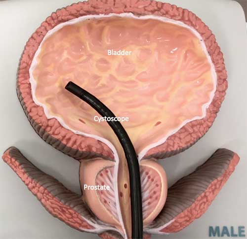
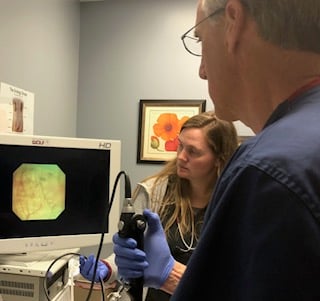
The lower part of the urinary tract, the bladder and urethra, is usually studied by cystoscopy.
Cystoscopy refers to the visual inspection of the bladder and urethra. This is carried out by inserting a small fiberoptic catheter into the urethra and the bladder which allows direct visualization of these structures. This is typically carried out under a local anesthetic in an examination room in the office.
Other tests may be done as well. A urine culture may be done to check for infection. Urinary cytology may help determine if there are any cancerous or pre-cancerous cells that are being shed into the urinary system. Cytology is carried out by doing a “Pap smear” on the voided urine.
Further Tests
In some patients, the above studies may prompt the need for further investigation which can include either an MRI study or retrograde pyelogram. An MRI uses a different type of imaging to give further information about the urinary system. A retrograde pyelogram is a study that can provide more detailed information about the ureters. If the ureters are not seen well or appear abnormal on the CT urogram or sonogram, then a retrograde pyelogram may give further details. A retrograde pyelogram study is carried out at the time of cystoscopy by inserting a small catheter into the ureter. Dye is injected through this catheter to demonstrate the ureter. This is typically carried out in the outpatient area of the hospital where the x-ray unit is available to make the necessary pictures.
In most cases, the finding on the CT urogram (or sonogram) and cystoscopy are normal and nothing further needs to be done. If there is an abnormality that is discovered, then it can be treated appropriately as indicated. Again, the main goal of evaluation is to make sure that the blood in the urine is not a sign of something which may pose a threat to the patient’s health. In the case where nothing serious is found, symptoms to watch for in the future which may indicate a sign of a problem include a change in the urinary pattern to more frequent urination, blood that can be seen in the urine, painful urination or pain that originates in the kidney. Kidney pain typically originates high in the back under the ribs and radiates down to the groin area. If these symptoms develop then repeat evaluation may be indicated.
Hematuria is a common problem and with the appropriate workup, the necessary treatment can be carried out.
We are pleased to offer online appointment request! New patients can request a call to schedule an appointment. Established patients seen in the last 3 years can request an appointment with one of our nurse practitioners/physician assistants.