

UPJ Obstruction
Ureteropelvic junction obstruction (UPJO) is a term used to describe blockage that involves the upper part of the urinary drainage system. To understand UPJO, it helps to begin with a description of the urinary system. The kidneys, one on each side, sit high in the upper abdomen towards the back.
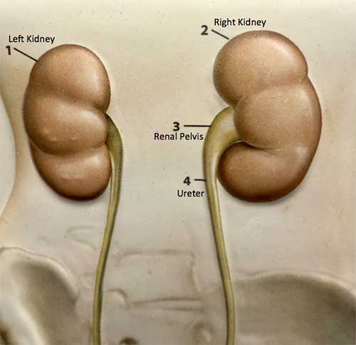
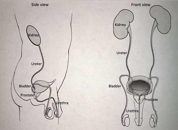
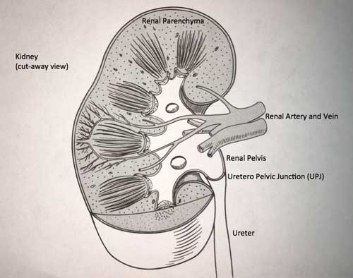
The kidneys filter the blood to extract excess fluid and waste products to form the urine. Urine, once made by the substance of the kidney (called the renal parenchyma) drains into the first part of the urinary collecting system called the renal pelvis. The renal pelvis then drains into the ureter, which is a long, thin tube that transports the urine to the bladder. The bladder stores urine until it is full and then empties to the outside during urination through the urethra.
UPJO occurs when there is blockage at the junction of the renal pelvis and the ureter. The blockage may be due to an anatomic reason, such as a blood vessel to the kidney crossing over top of the ureter at the ureteropelvic junction, or it may be due to a narrowed and nonfunctioning segment of the ureter.
How does UPJ obstruction present?
UPJO may show itself in a variety of ways. It may cause pain, infection, stone, or hematuria (blood in the urine). At times, it may not cause any symptoms, but may be detected during some type of imaging study (e.g., CT scan, ultrasound, MRI) done for evaluation of some other problem. If there is pain, it may be intermittent in nature. Pain develops within the urinary tract when the kidney is blocked and under pressure. In some cases, obstruction of the UPJ may be intermittent so pain may only develop during times of increased urinary output by the kidney, which can occur following a period of increased fluid intake or may occur after exposure to a diuretic substance, which can include coffee, tea, or alcohol.
How is UPJ obstruction evaluated?
Several tests are available to evaluate UPJO. A renal sonogram may show a dilated renal pelvis (called hydronephrosis or pelvicaliectasis on an imaging report). A CT scan may show the same, and can be useful to exclude other reasons for blockage, including extrinsic compression (something outside the collecting system pushing on it) or intrinsic blockage (something inside the collecting system causing blockage, such as a stone or a tumor). When UPJO is identified on an ultrasound or CT, the renal pelvis is dilated but the ureter below the point of blockage is not dilated.
Both the ultrasound and CT may allow evaluation of the thickness of the renal parenchyma, which may provide insight into the quality of the affected kidney. In the presence of long-standing, high grade obstruction, the back pressure on the kidney may have eroded away the substance of the kidney, so there is not much parenchyma remaining suggesting the kidney does not have much function.
A retrograde pyelogram can provide a detailed picture of the anatomy of UPJO. A retrograde pyelogram, which is an outpatient procedure done under anesthesia, is performed by the urologist who looks into the bladder with a fiber optic instrument (a procedure known as cystoscopy) which then permits injection of dye into the ureter through a catheter while watching with x-ray imaging.
A renal scan allows assessment of the degree of function of the involved kidney, and can quantify the affected kidney’s contribution to the overall level of function by both kidneys. A renal scan may also quantify the degree of obstruction by measuring the time it takes for the kidney to drain to the bladder.
How is UPJ obstruction treated?
Once UPJ has been assessed, there are a variety of treatment options available. If the UPJO is asymptomatic (if, not causing any symptoms) and there is good preservation of function, the affected kidney may be left alone with plans for follow-up monitoring. Similarly, if the UPJO is a symptomatic and the involved kidney has little to no function, with the recognition that the other kidney is doing most of the work, the involved kidney can be left as is.
When there is infection, stone, pain, or impaired function in an otherwise worthwhile kidney, then intervention is indicated. A variety of surgical techniques are available to address UPJO. For years, the standard approach has been open surgery carried out through an incision in the side of the abdomen (flank). The narrowed segment of the ureter is removed and the renal pelvis and ureter are joined back together (this procedure is known as a dismembered pyeloplasty).
In more recent years, the same procedure can be carried out laparoscopically, which may include the use of the DaVinci surgical robot, a tool for advanced laparoscopic technique.
Endoscopic approaches to UPJO are also available. A scope can be advanced through the bladder and the ureter (ureteroscopy) up to the narrowed segment which can then be incised (this procedure is called a retrograde endopyelotomy).
A scope may also be advanced through a small puncture site in the back into the kidney and renal pelvis (percutaneous nephrostomy) to approach the narrowed segment and incise the blockage (this procedure is called an antegrade endopyelotomy).
With any of these procedures, a stent is placed. A stent is a thin plastic tube, like a straw, that goes from the renal pelvis, across the site of the repair, through the ureter and into the bladder. The stent usually stays in for a period up to six weeks until healing has taken placed.
In some cases, the affected kidney may have minimal or no function, and the best approach may be surgical removal of the kidney (nephrectomy).
What are the risks of UPJ obstruction repair?
The surgical efforts to help address UPJO may introduce hazards, which include the general risks of surgery, such as bleeding or infection, and, more specifically, may cause problems, such as damage to adjacent structures, difficulties with urinary leakage at the repair site, or residual obstruction which may be due to scarring. In some patients, the portion of the stent which is in the bladder may cause voiding bother, including urinary frequency and urgency.
In summary, UPJO is a common urologic condition. When surgical correction is needed, a variety of treatment options are available to help manage the condition.
Print PageContact us to request an appointment or ask a question. We're here for you.