
Extracorporeal Shockwave Lithotripsy (ESWL) Surgery
Kidney stones are a common problem. Our Kidney Stone newsletter provides a broad overview of the management of kidney stones.
The urinary tract begins with the kidneys. The kidneys, one on each side, sit high in the upper abdomen partially underneath the rib cage. They filter the blood to extract excess waste products and fluid to form the urine. Urine, once formed in the kidneys, travels through a tube on each side, the ureter, down to the bladder. Urine is constantly being made by the kidneys and transported through the ureters into the bladder. The bladder stores urine until full and then empties to the outside through the urethra. Stones may be present in the kidney or the ureter. Stones can cause trouble when they block the drainage of urine out of the kidney.
Extracorporeal Shock Wave Lithotripsy (ESWL) is a valuable technique available to help manage kidney stones. ESWL is a technique that uses shockwave energy to fragment a stone into smaller pieces so that these smaller pieces can be passed more easily.
The ESWL procedure is done in the operating room in the hospital or the surgery center. The procedure takes about 1 hour. Patients are given IV sedation or a general anesthetic. During the procedure the patient rests on the lithotripsy table. The patient is positioned so that the stone is in a precise treatment area, called the focal point, which is identified with the use of x-ray imaging.
Shockwaves generated by the lithotripsy machine travel through the body wall to reach the kidney stone. Shockwaves cause the kidney stone to break into smaller pieces.
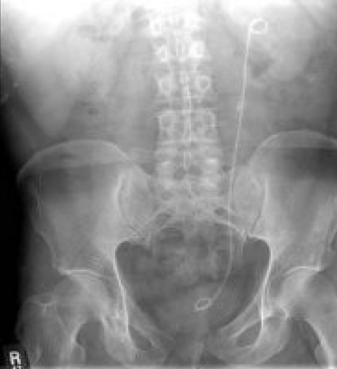
Patients are usually discharged several hours after the procedure. In some cases, as part of the ESWL procedure, a stent may be placed to promote passage of stone pieces or to prevent the kidney from becoming blocked. (See more details in Kidney Stone newsletter)
Postoperatively, patients may pass fragments over the next few weeks. There may be blood in the urine which may come and go. In some cases, the pieces pass without any difficulty. Other times, there may be “kidney stone pain” as the pieces work their way through. Pain pills can be used as needed for symptom relief (see Instructions for Passing Kidney Stone newsletter)
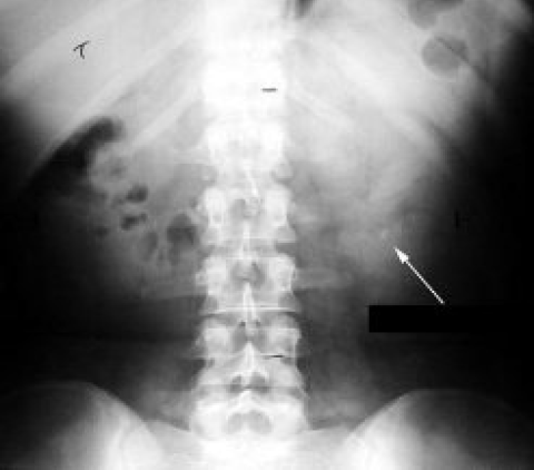
Office follow up is typically carried out several weeks after ESWL. Some type of imaging (for example, a KUB which is a plain abdominal x-ray) may be checked to assess for remaining stone pieces.
Stones break up differently depending on their crystalline structure. Some stones fragment easily into small bits of dust and there may be no remaining pieces. Other stones may have a harder crystalline structure and may break into larger size fragments which may remain, or the stone may not break up at all. In some cases, additional procedures may be needed which might include, repeat ESWL to address remaining fragments, or may require some other type of procedure such as ureteroscopy (see Kidney Stone newsletter).
ESWL is a common procedure performed on a regular basis by the physicians of The Urology Group and provides a useful technique to help patient suffering with kidney stones.
Print PageContact us to request an appointment or ask a question. We're here for you.